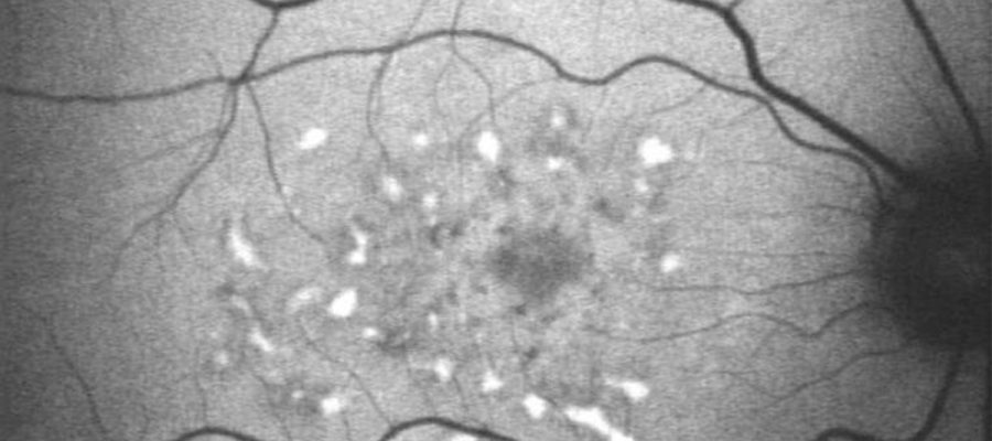
Fundus autofluorescence (FAF) imaging is a valuable noninvasive technique that has revolutionized the study of the retina and is still a very important tool on retina image because helps diagnosis and treatments to be more accurate. By mapping naturally or pathologically fluorophores in the ocular fundus, FAF provides ophthalmologists with a unique window into the metabolism of the retina.
The principle behind fluorescence involves the absorption of light at one wavelength and its re-emission at a longer wavelength, resulting in the generation of an image of distinct colors. Among the various fluorophores found in the fundus eye, lipofuscin stands out as one of the most significant. This intracellular pigment accumulates with cellular aging and oxidative stress, serving as a crucial marker for cellular changes.
By capturing the natural fluorescence of the eye’s inner tissues, FAF unveils a hidden world of cellular changes and metabolic activity. It’s like turning on a fluorescent light in a dark room, revealing intricate details that were previously unseen. This revelation holds immense clinical value, as it allows for the mapping of endogenous and pathological fluorophores in the ocular fundus, providing critical insights into various retinal diseases.
The importance of FAF cannot be overstated. Understanding these intricate changes enables early detection and diagnosis of conditions such as age-related macular degeneration, leading to better treatment outcomes and improved patient care.
To stay informed and keep up with the latest advancements in the field, we invite you to follow our social media channels and register on our blog. You can find the link in the bottom of this page.
This is ORBIT: science combined with the opinion of those with experience for you to use in practice, effectively and consistently!

